Scoliosis Patients

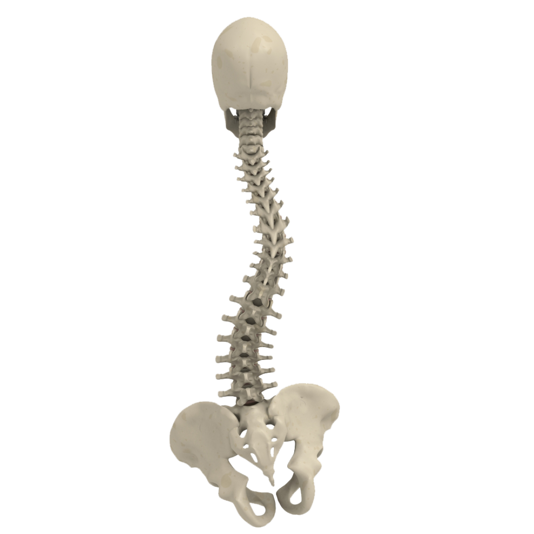
Scoliosis is a curvature of the spine by more than 10 degrees; typically in a “S” or “C” shape which can lead to poor posture, muscle weakness, pain, and even heart lung problems.
In more than 80% of the cases, a specific cause is not found and such cases are termed “idiopathic,” meaning “of undetermined cause.” It is by far the most common kind of scoliosis and it develops most frequently in preteens or teenagers. In adolescent idiopathic scoliosis (AIS), the primary surgery used today is fusion surgery.
The goal of is two-fold. First, to prevent curve progression and secondly to obtain some curve correction. However, during this procedure patients receive a high dose of radiation and many of them emerge from surgery with screws of concern and complications.
A Large Number of Patients Treated for Scoliosis Emerge from Surgery with Screws of Concern
Screws at risk impinging anatomic structures such as the aorta, esophagus, trachea or lung and screws causing neurological deficits or injuries are reported in two recent large studies1,2.
Risk of Vascular Complications
In patients with deformity, revision surgery to remove a screw at risk for the aorta is recommended in 4.7% (7/1483 and 6/1272) of the patients.
Radiation Exposure on Young Patients
– The radiation exposure is greater during scoliosis surgery8.
– Young people are very sensitive to radiation exposure during their childhood9.
– Girls are at a higher risk of cancer development10.
– Intra-operative navigation increases the radiation exposure to patients especially in AIS; When accounting for clinical and radiographic differences, patients treated with intraoperative navigation received 8.18 mSv more radiation. Authors project that intraoperative navigation will generate approximately one iatrogenic malignancy for every 1,000 AIS patients treated11.
Instability
Misplaced screws without clinical symptoms can lead to screw loosening (1.4%: 3/208 patients4 ; 3.5%, 3/86 patients5).
Revision Surgery
Pedicle screw misplacement is the first cause of reoperation within 30 days of spine surgery in 1.7% AIS a patients in a US multi center study7.
Cerebrospinal Fluid (CSF) Leaks
– CSF leakage without clinical symptoms from screw holes in 3.5% (3/86) patients5.
– Dural leak causing positional headaches in 0.9% (3/322) patients6.
How DSG Technology Can Help
A randomized clinical trial12 assessed the accuracy and time for pedicle screw placement with the DSG Technology and the Free-Hand technique for posterior scoliosis surgery.
- There were totally 3 times less pedicle perforations in the DSG group (4.1%) than in the Free-Hand group (14.2%) (15 versus 47 (P<0.001)).
- The average screw insertion time was decreased by 15% in the DSG group.
A retrospective study13 in patients with severe deformities showed a greater screw placement accuracy in the DSG Group compared with the Free-Hand technique, especially in the thoracic region (92.5% DSG Group vs 87% FH group).A retrospective study14 in patients with severe syndromic and neuromuscular scoliosis, demonstrated a 10.2% higher screw placement accuracy when using PediGuard and twice lower abandonment rate due to liquorrhea or perforation.
….the PediGuard can reduce exposure to fluoroscopy, has high sensitivity and specificity for detecting pedicle perforations, and can significantly reduce the number of malpositioned screws
NICE 201515
References
1 – Makhni MC et al. Spine J. 2017 Jun 30. pii: S1529-9430(17)30312-1. doi: 10.1016/j.spinee.2017.06.038. [Epub ahead of print]
2 – Sarwahi V et al. Spine (Phila Pa 1976). Spine (Phila Pa 1976). 2016 May;41(9):E548-55.
3 – Sarlak A et al. Eur Spine J. 2009 Dec;18(12):1892-7. Epub 2009 Jun 14.
4 – Li G et al. Eur Spine J. 2010 Sep;19(9):1576-84.
5 – Sugawara R et al. Eur Spine J. 2015 Mar 8. [Epub ahead of print]
6 – Floccari LV. J Pediatr Orthop. 2017 May 17.[Epub ahead of print]
7 – Samdani AF et al. Spine. 2013 Oct 1.
8 – O’Donnell C et al. Spine (Phila Pa 1976). 2014 Jun 15;39(14):E850-5.
9 – Luan FJ et al. European Spine Journal (2020) 29:3123–3134
10 – Miglioretti DL et al. 2013 Aug 1;167(8):700-7. doi: 10.1001/jamapediatrics.2013.311
11 – Striano Spine J. 2024 Jan 21:S1529-9430(24)00018-4. doi: 10.1016/j.spinee.2024.01.007.
12 – Bai YS et al. 2013 Aug;26(6):316-20.
13 – Allaoui et al. (2018 Dec;120:e466-e471. doi: 10.1016/j.wneu.2018.08.105. Epub 2018 Aug 24.)
14 – Yurube et al. J. Clin. Med. 2022, 11, 419. https://doi.org/10.3390/jcm11020419
15 – NICE. Medtech innovation briefing. The PediGuard for placing pedicle screws in spinal surgery. Published: 25 March 2015
Anterior Scoliosis Correction
Anterior Scoliosis Correction provides an important alternative to treat spinal deformity
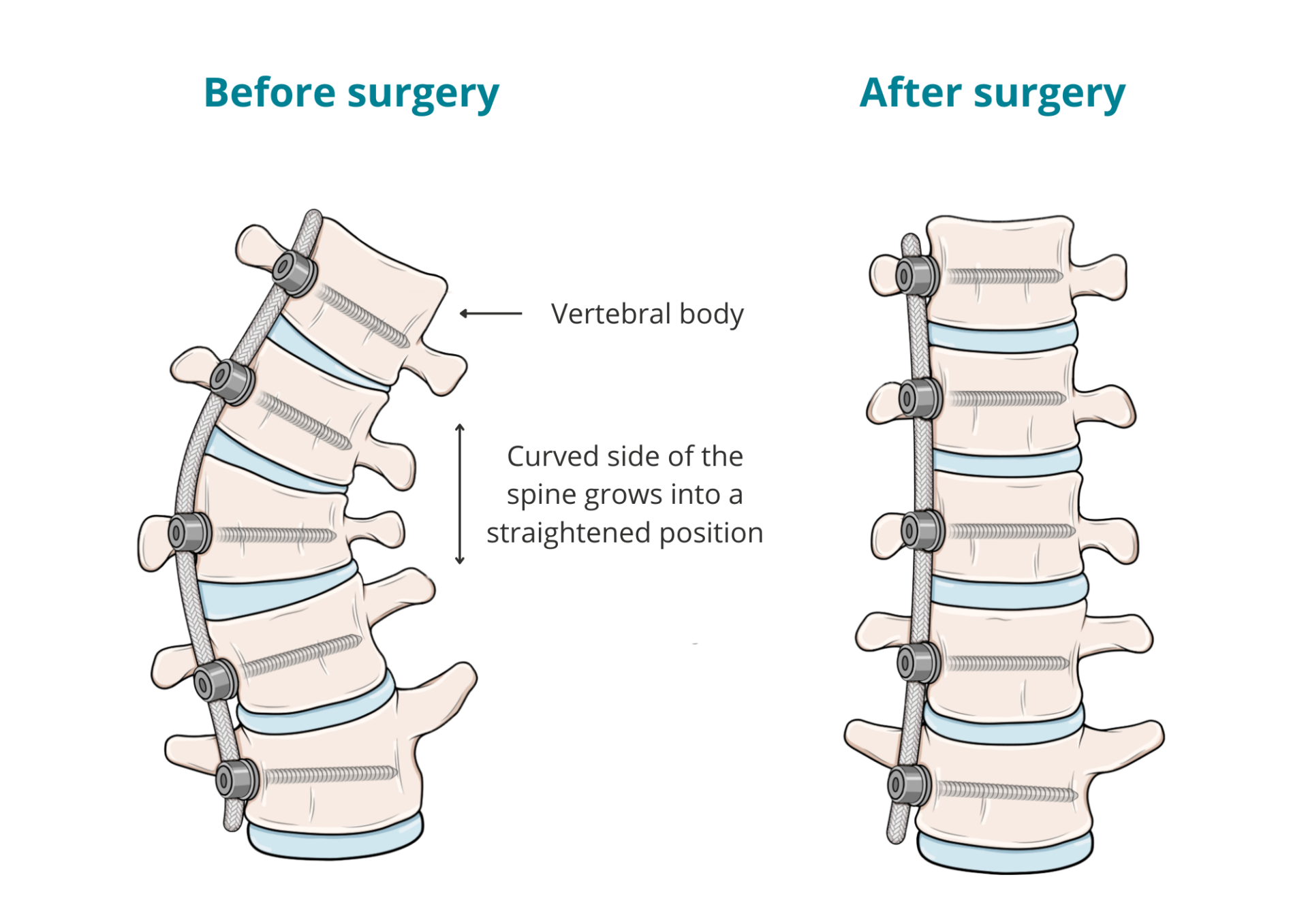
It is a minimally invasive procedure performed by most surgeons through thoracoscopy or mini-open thoracotomy to allow maintenance of spine mobility and shorter recovery time. Specific medical implants have been developed for anterior scoliosis correction and are composed of screws that are placed on the lateral part of each vertebral body, on the convex side of the curve, and a single synthetic tether spans between the screws. The lungs are selectively ventilated allowing the lung on the convex side to collapse creating the working space. Tension is then applied to the tether to obtain a peri-operative correction, which is equivalent to a “brace-effect”. The correction improves over time due to a “convex side epiphysiodesis effect” if the patient has significant remaining growth1.

Challenges In Anterior Scoliosis Correction
A significant Amount of X-Ray Shots is Necessary to Control Screw Placement
Anterior Scoliosis Correction presents new challenges in respect to radiation exposure, as screws cannot be placed free-hand and the lateral positioning of the patients increases scattered radiation.
The use of PediGuard threaded can effectively reduce radiation exposure up to 41%2 (from 9.16 to 5.52 mGy*m2 per screw overall).
Screw Length Determination is crucial
In the case of Anterior Scoliosis Correction, bicortical screws are inserted in the lateral, convex aspect of the vertebral body.
While the entry point and trajectory of the screw are determined by anatomical landmarks, the PediGuard alerts the surgeon that the cortical bone on the concave side has been breached without necessity of subsequent X-rays. The laser marks every 2.5 mm help assess insertion length into the bone.

The use of the Threaded PediGuard device provides a real time feedback that help the surgeon to overcome theses challenges.
References
1 – Courvoisier, Vertebral body tethering in AIS Management – A preliminary report. Children 2023, 10, 192. https://doi.org/10.3390/children10020192
2 – Da Paz, S., Trobisch, P. & Baroncini, A. The use of electronic conductivity devices can effectively reduce radiation exposure in vertebral body tethering. Eur Spine J 32, 634–638 (2023). https://doi.org/10.1007/s00586-022-07489-0
Vous avez des questions ?
